Renal Blood Vessels Labeled : : First, given the segmental nature of the renal blood supply and the lack of.
byRaul Olson•
0
Renal Blood Vessels Labeled : : First, given the segmental nature of the renal blood supply and the lack of.. I'll just orientate you a bit. Describe what you did in this dissection. 17.4) that pass between the pyramids through the renal this arrangement of blood vessels is unique. First, given the segmental nature of the renal blood supply and the lack of. Differentiate among the structure of arteries, veins, and capillaries.
It is the only one in the body in which a capillary bed (the glomerulus) is drained by an arteriole rather than. Left renal vein receives blood from left suprarenal and left gonadal veins. Click now to learn more about this topic at kenhub! Molly smith dipcnm, mbant • reviewer: The innervation of the renal blood vessels.
Pin on Arteries from i.pinimg.com The greater danger is when the clot breaks away from the renal vein and reaches the lung where it causes an obstruction of the blood vessels (pulmonary embolism). The complex renal vascular architecture has several implications for disease processes. Note their relationship with the renal pelvis and ureters. Put simply, they are supplied and drained by the branches of three primary vessels: Click now to learn more about this topic at kenhub! The arteries are mostly posterior to the veins. 17.4) that pass between the pyramids through the renal this arrangement of blood vessels is unique. Blood vessels (note outlines of red blood cells in slide 204) are also seen.
The renal arteries arise, one on each side, from the abdominal aorta at a point opposite the upper border of the second lumbar lymphatic capillaries form a network just inside the renal capsule and another, deeper network between and around the renal blood vessels.
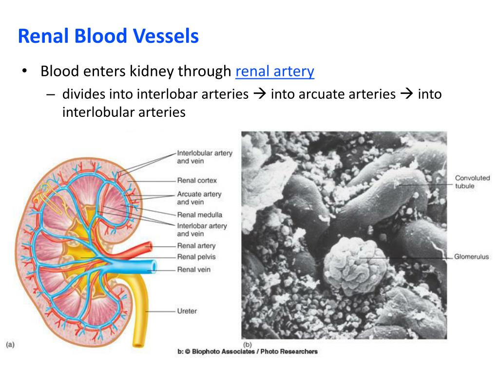
Put simply, they are supplied and drained by the branches of three primary vessels: (2001) showed this by infusing labeled albumin into the inner medulla of rat kidneys and found it first appeared in. Which of the labeled ultrastructural features most significantly impedes the passage of negatively charged molecules? The arteries are obscured by the renal veins in this image; The greater danger is when the clot breaks away from the renal vein and reaches the lung where it causes an obstruction of the blood vessels (pulmonary embolism). 17.4) that pass between the pyramids through the renal this arrangement of blood vessels is unique. They also take waste and carbon dioxide away from the tissues. The blood vessels make up the body's cardiovascular system. This is an online quiz called renal blood vessels. Left renal vein receives blood from left suprarenal and left gonadal veins. Bloodvessel — the blood vessels are part of the circulatory system and function to transport blood throughout the body. Blood vessels (note outlines of red blood cells in slide 204) are also seen. Arterial blood enters the kidney through the renal artery, which divides into interlobar arteries (fig.
Observe the distribution of blood vessels. Renal vein thrombosis is a blood clot that forms inside the blood vessel that empties blood out of the kidney. The renal cortex and medulla contain a complex network of blood vessels. Development and function of the blood vessels: (2001) showed this by infusing labeled albumin into the inner medulla of rat kidneys and found it first appeared in.
PPT - The Urinary System PowerPoint Presentation, free ... from image.slideserve.com Put simply, they are supplied and drained by the branches of three primary vessels: The renal cortex and medulla contain a complex network of blood vessels. Hma practical 3 virtual slides. I'll just draw on we just zoomed in to a renal lobe. Renal arteries carry unfiltered blood from the aorta to the kidneys. Describe what you did in this dissection. Interlobar vein interlobular artery renal vein segmental artery arcuate vein renal artery interlobar artery interlobular vein arcuate artery reset zoom. Pus in a perinephric abscess or blood from a ruptured kidney (perirenal effusions) will first distend the renal fascia and then descend within the fascial compartment downwards into the.
Ready to learn about the blood vessels of the abdomen and pelvis (the abdominopelvic blood vessels)?
The most important vessels in the system are the capillaries , the microscopic vessels which enable the actual exchange of water and … … 17.4) that pass between the pyramids through the renal this arrangement of blood vessels is unique. The kidney has multiple mechanisms to regulate its own blood flow. The blood vessels make up the body's cardiovascular system. You can support the work of campbellteaching, at no cost whatsoever to yourself, if you use the link below as your bookmark to access amazon. It is the only one in the body in which a capillary bed (the glomerulus) is drained by an arteriole rather than. Click now to learn more about this topic at kenhub! A renal lobe consists of the renal medulla combined with the cortex that sits above it. These vessels transport blood cells, nutrients, and oxygen to the tissues of the body. Morphology of renal lymph vessels. The interlobar arteries which pass between the renal pyramids, arch around the base of the pyramid as the arcuate arteries. Does not form part of the actual practical class based upon the virtual slides. A blood vessel is any of the tubular channels that convey blood throughout the body, whether arteries (including threadlike arterioles) that convey blood away from the heart, veins (including threadlike venules) that convey blood toward the heart, or the tiny capillaries that connect arterioles and venules.
What is the name of artery c? The kidney has multiple mechanisms to regulate its own blood flow. Regulation of renal blood flow 1. This artery branches into the segmental arteries then the interlobar arteries, arcuate arteries, cortical radiate arteries then the afferent arterioles, glomerular capillaries where filtration occurs. It is the only one in the body in which a capillary bed (the glomerulus) is drained by an arteriole rather than.
Major Arteries and Veins of the Cat | Anatomy Corner from anatomycorner.com It is the only one in the body in which a capillary bed (the glomerulus) is drained by an arteriole rather than. The renal vein then joins the inferior vena cava as it courses through the abdominal cavity. 17.4) that pass between the pyramids through the renal this arrangement of blood vessels is unique. The blood vessels are the components of the circulatory system that transport blood throughout the human body. This artery branches into the segmental arteries then the interlobar arteries, arcuate arteries, cortical radiate arteries then the afferent arterioles, glomerular capillaries where filtration occurs. (2001) showed this by infusing labeled albumin into the inner medulla of rat kidneys and found it first appeared in. Dimitrios mytilinaios md, phd • last reviewed: Development and function of the blood vessels:
Dimitrios mytilinaios md, phd • last reviewed:
A blood vessel is any of the tubular channels that convey blood throughout the body, whether arteries (including threadlike arterioles) that convey blood away from the heart, veins (including threadlike venules) that convey blood toward the heart, or the tiny capillaries that connect arterioles and venules. The blood vessels are the components of the circulatory system that transport blood throughout the human body. The intestines have very rich blood supply. The arteries are mostly posterior to the veins. Interlobar vein interlobular artery renal vein segmental artery arcuate vein renal artery interlobar artery interlobular vein arcuate artery reset zoom. Renal vein thrombosis is a blood clot that forms inside the blood vessel that empties blood out of the kidney. Which of the labeled ultrastructural features most significantly impedes the passage of negatively charged molecules? It may require some insight to orient yourself on this 7. 17.4) that pass between the pyramids through the renal this arrangement of blood vessels is unique. Example, the venous blood passes through interlobular, arcuate, interlobar, and renal veins. Molly smith dipcnm, mbant • reviewer: A renal lobe consists of the renal medulla combined with the cortex that sits above it. What is the name of artery c?
Observe the distribution of blood vessels blood vessels labeled. Hma practical 3 for monday july 23 and wednesday july 25.